Cranial Nerves
Optic nerve - I : vision
Olfactory nerve - II smell
Oculomotor nerve - III eye ball movement
Trochlear nerve - IV eye ball movement
Trigeminal nerve - V facial sensation
Abducent nerve - VI eye ball movement
Facial nerve - VII facial expression & taste in anterior 2/3 of tongue
Vestibula auditory nerve - VIII Balance, hearing
Glossopharyngeal nerve - IX Sensation of throat and part of tongue
Vagus nerve - X Internal organs
Accessory nerve - XI Shoulder
Hypoglossal nerve - XII Movement of the tongue
Facial Nerve
Seventh cranial nerve.
Mixed nerve (sensory and motor)
Called the nerve of facial expression
Origin
It emerges from the lower border of pons as two roots, large medial motor root and a small lateral sensory root (nervus intermedius)
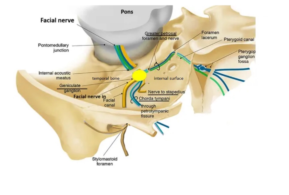
Course
Leaves the cranial cavity by passing through the internal acoustic meatus which is an 1cm long opening in the petrous part of the temporal bone
It then enters the facial canal which is divided into three segments namely labyrinthine, tympanic and mastoid
Within the facial canal, three important events occur:
Firstly the two roots fuse to form the facial nerve.
Next, the nerve forms the geniculate ganglion (a ganglion is a collection of nerve cell bodies).
Lastly, the nerve gives rise to:
Greater petrosal nerve parasympathetic fibres to mucous glands and lacrimal gland.
Nerve to stapedius motor fibres to stapedius muscle of the middle ear.
Chorda tympani special sensory fibres to the anterior 2/3 tongue and parasympathetic fibres to the submandibular and sublingual glands.
The facial nerve then exits the facial canal (and the cranium) via the stylomastoid foramen. This is an exit located just posterior to the styloid process of the temporal bone.
Labyrinthine segment :
Facial canal lies above the vestibule of internal ear.
The facial nerve passes through this part to reach the medial wall of middle ear where it bends (external genu - location of geniculate ganglion sharply backwards.
Tympanic segment :
The nerve then passes through the tympanic segment of facial canal which runs horzontally backward along the medial wall of the middle ear till it reaches the junction of medial and posterior wall of middle ear.
Mastoid part :
the nerve passes vertically downward in the mastoid part of facial canal in the posterior wall of middle ear to reach the stylomastoid foramen
After emerging from the stylomastoid foramen the facial nerve turns superiorlyand runs forwards crosses the styloid process and enters the posteromedial surface of parotid gland just anterior to the outer ear
The first extracranial branch, the posterior auricular nerve provides motor innervation to the some of the muscles around the ear, the posterior belly of the digastric muscle and to the stylohyoid muscle.
Immediately distal to this, motor branches are sent to the posterior belly of the digastric muscle and to the stylohyoid muscle.
The main trunk of the nerve, now termed the motor root of the facial nerve, continues anteriorly and inferiorly into the parotid gland (Note : parotid gland is innervated by the glossopharyngeal nerve).
Within the gland it lies lateral to retromandibular vein and external carotid artery
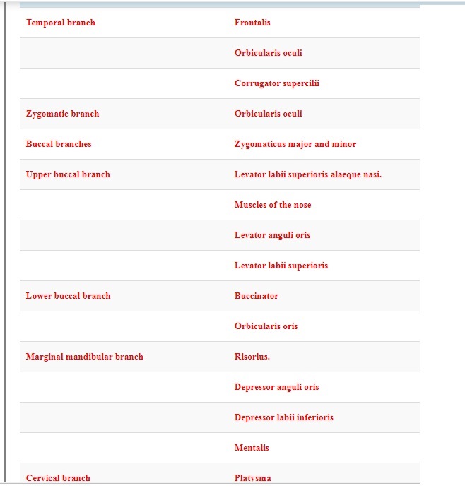
It divides into 5 terminal branches viz. temporal, zygomatic, buccal, marginal mandibular and cervical
These branches are responsible for innervating the muscles of facial expression.
Special Sensory Functions
The chorda tympani branch innervates the anterior 2/3 of the tongue - special sense of taste.
The nerve arises in the facial canal, and travels across the bones of the middle ear entering the infratemporal fossa. Here, the chorda tympani hitchhikes with the lingual nerve. The parasympathetic fibres of the chorda tympani stay with the lingual nerve, but the main body of the nerve leaves to innervate the anterior 2/3 of the tongue.
Parasympathetic Functions
The parasympathetic fibres of the facial nerve are carried by the greater petrosal and chorda tympani branches.
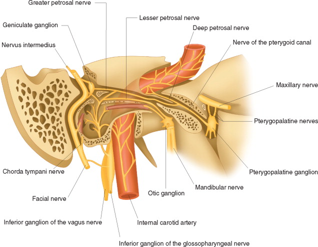
Greater Petrosal Nerve
The greater petrosal nerve arises immediately distal to the geniculate ganglion within the facial canal. combining with the deep petrosal nerve to form the nerve of the pterygoid canal ; enters the pterygopalatine fossa and synapses with the pterygopalatine ganglion ; branches from ths ganglion provide parasympathetic innervation to the mucous glands of the oral cavity, nose and pharynx, and the lacrimal gland.
Chorda Tympani
The chorda tympani also carries some parasympathetic fibres. These combine with the lingual nerve (a branch of the trigeminal nerve) in the infratemporal fossa and form the submandibular ganglion. Branches from this ganglion supply submandibular and sublingual salivary glands.
Applied Aspects
Bell's Palsy - lower motor neurone (LMN) paralysis
UMN (upper motor neuron) lesion results in paralysis of the contralateral middle and lower parts of the face with sparing of the muscles of the forehead and the orbicularis oculi muscle.
* * * * * * * *
University question :
Name the cranial nerves. Describe the facial nerve under the following headings
Course and distribution
Branches
Applied aspects
.
. Name the cranial nerves. Describe the facial nerve under the following headings
Course and distribution
Branches
Applied aspects